Authors
Michelle S. Swedek, BS1, Alexander Hall, MS1, Tami DenOtter, MD1
1Creighton University School of Medicine, Omaha, NE
Conflict of Interest Statement
The authors declare no conflict of interest.
Corresponding author
Brief Description
Diagnostic radiology is a diverse medical specialty essential to effective and efficient diagnosis and treatment. To promote interest and awareness in this specialty, medical students should have the opportunity to explore an engaging radiology curriculum and gain fundamental skills. Historically, radiology education in the undergraduate medical setting has been limited due to challenges in providing hands-on learning and stimulating critical thinking (O’Connor et al., 2016). To address these obstacles, we designed a new radiology simulation lab as part of the radiology elective curriculum for medical students. One year after the lab’s implementation, medical students who participated were surveyed regarding the lab. Overall, medical students positively ranked the lab’s structure, education, timeline, and enjoyment. These results highlight the value of an interactive radiology curriculum in undergraduate medical education.
Introduction
Diagnostic radiology is a diverse medical specialty essential to effective and efficient diagnosis and treatment. A successful radiologist must have attention to detail, the ability to work in fast-paced and stressful situations, and strong critical thinking skills (Collins et al., 2002). Medical students and radiologists from across the United States argue that radiology needs to have a greater role in medical education for all medical students, regardless of career ambitions (Dmytriw et al., 2015; Gunderman et al., 2003; Zwaan et al., 2017).
Given these calls to action and the unique skillset of radiologists, medical students should have the opportunity to explore an engaging radiology curriculum and gain fundamental skills. One challenge with radiology education is providing hands-on learning and stimulating critical thinking (O’Connor et al., 2016). Radiologists typically work alone, and the traditional observational model may not be ideal for students and faculty. A shadowing student may lose focus after watching the radiologist interpret many images, while a radiologist may not have adequate time to review images with the student during a busy day (Redmond et al., 2020). In this model, students may not be exposed to classic radiology cases and are not actively engaged in reading images. This may lead to decreased interest in the field and greater uncertainty in interpreting radiological images (Redmond et al., 2020).
To address these challenges, institutions across the country have implemented flipped classroom and simulation-based programs that have successfully enhanced learner knowledge, engagement, and interest in radiology (Belfi et al., 2015). At Creighton University School of Medicine, a new radiology simulation lab has been added to the radiology elective for medical students. The radiology simulation lab is an automated course featuring introductory and instructional videos, hands-on case viewing, and post-educational radiology read-out sessions for ten radiology subspecialties. The course is divided into the following subspecialties: chest, advanced cardiothoracic, gastrointestinal, genitourinary, neurosciences/neuroradiology, orthopedic/musculoskeletal, women’s, interventional radiology, nuclear medicine/oncology, and pediatric imaging.
The simulation course is divided into subspecialties to teach level-appropriate radiology topics for optimizing patient care and understanding at three educational levels: medical students, mid-level providers (such as physician assistants and advanced practice nurses), and non-radiology residents. This radiology simulation course offers radiology content specific to various specialties and accessible to learners at different educational levels. Each subspecialty is expected to take approximately two to three hours to complete. The ten subspecialties contain two to six different radiology educational teaching topics, each consisting of an instructional video, radiology case viewing, and post radiology read-out session.
The instructional videos cover fundamental radiology topics and the interpretation of various diagnoses or differential diagnoses within the subspecialty. The educational topics are based on the Association of University Radiologists Alliance of Medical Student Educators in Radiology Curriculum, Competencies and Learning Objectives for Medical Students (AMSER Curriculum, Competencies, and Learning Objectives, 2023). These radiology topics parallel Radiology Aquifer Cases and can function as stand-alone or supplementary teaching tools to Radiology Aquifer Case Modules (Aquifer Radiology, 2024).
At our institution, the simulation lab is conducted as an in-person learning activity with small groups of three to four students. The course is a ten-day elective, with one day dedicated to each radiology subspecialty. Each day, the students begin one of the modules with a 10minute teaching video before working as a group to view and diagnose the accompanying radiology cases. For example, on the neuroradiology day, there are five different education topics/modules: Approach to Head Trauma, Early Signs of an Acute Stroke, Cervical Spine Fractures, Epidural Hematoma, and Subdural Hematoma. Within these five educational topics, there are multiple case examples and a single post read-out session. The read-out video features a radiologist interpreting the case, highlighting the approach to reading the radiology exam, and identifying key radiologic findings. This allows students to verify their findings and interpretation of the images.
Approximately 50-60 educational topics and 400 student-level radiology cases (including radiographs, ultrasound, CT, MRI, PET imaging, mammography, and interventional procedures) have been chosen for medical students, allied health students, and non-radiology residents.
These topics are organized in the ten subspecialities mentioned previously.
The cases are on a Picture Archiving and Communication System (PACS) driven workstation to simulate the work of a healthcare professional viewing the case. Horos PACS system is a free, open-source medical image viewer based on OsirX (Pixmeo SARL, Geneva, Switzerland), designed specifically for storing and viewing for medical imaging. Horos PACS allows students to launch, view, and manipulate images as a radiologist would when interpreting radiology exams. Since Horos is a free software program and the main operational component for running a radiology simulation lab, other institutions can easily develop and maintain similar labs. After downloading Horos, faculty can select appropriate images, deidentify them to remove patient information, and upload them to Horos PACS for viewing.
The students complete one of the ten radiology subspecialties sections each day of their elective. They are introduced to radiographs, computer tomography, MRI, PET imaging, nuclear medicine, and mammography. All images were selected from patients at our institution and chosen by a subspecialized attending radiologist to best exemplify their teaching topics. All images were de-identified and given unique a unique four-digit radiology simulation lab case number. Case numbers were assigned and grouped by radiology subspecialty. For multiple images of the same patient, the case numbers share the same first three digits, while the last digit is unique to each image.
The radiology simulation lab was recently implemented into the radiology elective curriculum, so we aimed to evaluate student perspectives of the lab. In this study, we surveyed fourth-year medical students who completed the lab during their radiology elective. We aimed to better understand where the new curriculum is successful and where it could be improved, especially regarding the structure, education, and time spent for the simulation lab.
Methods
Fourth-year medical students at Creighton University School of Medicine who participated in the radiology simulation lab from June 2022 to March 2023 were invited via email to complete the survey. The survey consisted of several Likert-scale multiple-choice questions. For each question, participants ranked their degree of agreement with a statement as follows: 1 = Strongly disagree, 2 = Somewhat disagree, 3 = Neutral, 4 = Somewhat agree, and 5 = Strongly agree. The survey was voluntary and anonymous to encourage honest and constructive feedback. To analyze the data, 1-sample t-tests were used to compare the sample average against a population value of 3, which is equivalent to neutral. A p-value less than 0.05 was considered statistically significant. The 95% confidence intervals (CI) were drawn from bootstrapping with 1000 samples.
Results
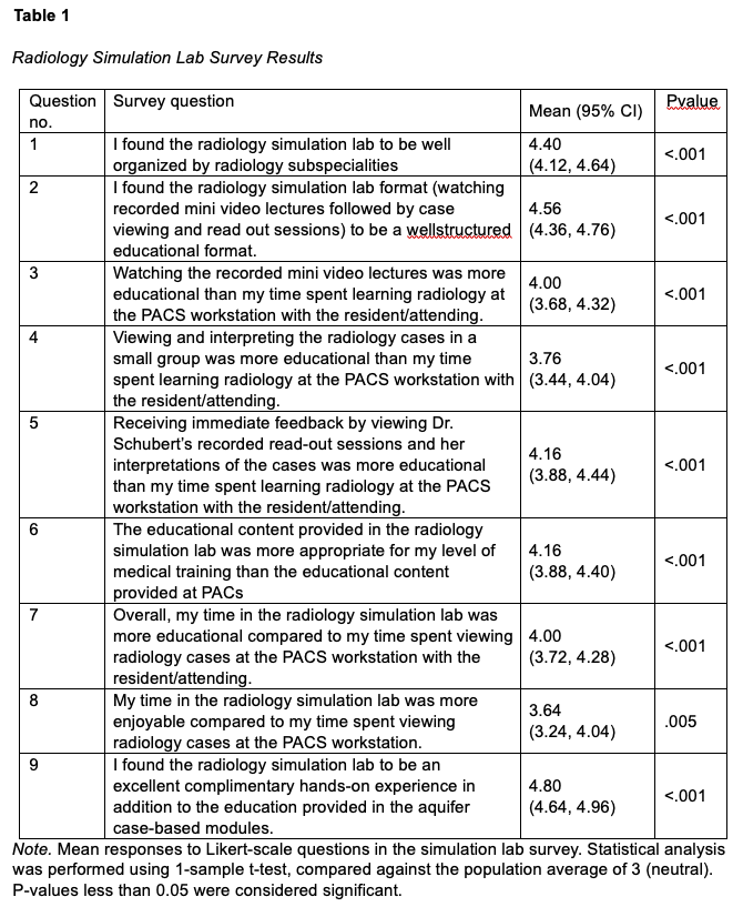
Thirty-nine students participated in the new simulation lab and were invited to complete the survey. Of those, 25 medical students responded, resulting in a 64% response rate. The survey questions, average response, 95% CI, and p-value are described in more detail in Table 1. All items evaluating simulation lab structure, education, timeline, and enjoyment received ratings higher than neutral (3), and these differences were statistically significant. Students agreed they learned more about subspecialties as evidenced by question 1. They also became more confident in their ability to interpret images (questions 2-5) and practice their clinical decision-making skills independently (question 4).
Discussion
The radiology clerkship aims to give medical students insight into how radiologists work and fundamental image interpretation skills. In the traditional model of radiology education, this proves challenging as students are only given passive learning opportunities, such as shadowing or listening to didactic lectures. With the rise of electronic learning in radiology, we now have the capability for students to immerse themselves in radiology through simulation (Zafar et al., 2014).
Overall, the students at our institution had a positive experience learning in the simulation lab as they combined traditional classroom learning with an interactive simulation lab. This is consistent with prior work demonstrating medical students preferred an active learning environment as opposed to a passive one in radiology education (Zou et al., 2011). Moreover, students perceived they were able to learn better in the simulation lab. Other institutions that implemented similar flipped classrooms found that students who participated performed better on competency exams compared to those learning through a traditional classroom (Friedman et al., 2017). Students also enjoyed learning in the radiology simulation lab more than shadowing. An educational model where students enjoy the learning experience and are engaged may help better solidify concepts and provide valuable exposure to radiology.
Other researchers in the field have recently encouraged the use of similar case-based models for third- and fourth-year medical students in their radiology elective (Farmakis et al., 2023).
This initial research project aimed to gather insight and early feedback from the students on their perception of the radiology simulation lab as a novel educational tool. We focused on the students’ enjoyment of the elective and their subjective education benefit. Future research should objectively assess the educational impact. For instance, a pretest of students’ interpretation of images could be compared to a posttest evaluation at the end of the two-week elective.
Conclusion
Our results indicate medical students had a positive learning experience in the new radiology simulation lab at our institution. They positively ranked the design, content, scheduling, and satisfaction of the lab. This study highlights the feasibility of a simulation-based radiology curriculum, and we call for a more interactive and engaging radiology education model.
References
AMSER Curriculum, Competencies, and Learning Objectives. (2023). Association of University Radiologists.